
The orthodontic report (portfolio)
is a standard set of 16 views extracted from the CBCT. We send it to you in the e-mail as separate 16 jpeg files. You will always have the information at your fingertips on any device with an e-mail access.
The composition of the portfolio is a compilation of known CBCT evaluation methods, the result of a long teaching of the method's use in orthodontic practice by orthodontist Larisa Korsak.
Cephalometry
right side view
Cephalometry
frontal view
TMJ
medial pole, X-ray view
TMJ
lateral pole, "Bone" view
Coronal slice view of the molar area
for the inclination of the lateral group of teeth evaluation
Palatal suture
done for patients younger than 18 years
Sequential full-volume views
profile right side
Sequential full-volume views
¾ profile right side
Sequential full-volume views
full face
Sequential full-volume views
¾ profile left side
Sequential full-volume views
profile left side
Airway measurement
Panorama
x-ray view without overlaying other anatomical structures
Panorama
"bone" view
Ceph
lateral
Ceph
straight
Symmetry analysis
The portfolio does not include any opinion or judgment of the physician about what is depicted in the jpeg file. We do not give any comments on the essence of the process or on the diagnosis based on portfolio data. This service exclusively provides a series of images in jpeg file format (portfolio) of various reformats and renderings highlighted in the CBCT viewer.
I WANT ORTHOREPORT
You can also write to us by e-mail to ortoreport@mail.ru
Clicking the button you confirm that you accept our confidentiality policy and allow us to process your personal data.
Clicking the button you confirm that you accept our confidentiality policy and allow us to process your personal data.
Processing time –
3 working days
3 working days
Price –
50 US Dollars
50 US Dollars
Creation of cephanalysis according to the doctor's individual requirements – 70 US Dollars (you pay once – use it indefinitely).
Processing time –
Creation of cephanalysis according to the doctor's individual requirements – 70 US Dollars (you pay once – use it indefinitely).
3 working days
Price –
50 US Dollars
PAYMENT
Price is 50 USD per report. Payment via PayPal for each report via link we send you after we get the scan from you.
HOW TO ORDER
The radiologist makes a CBCT scan according to the instructions
We send you a link to pay online
We make the report and send it to you
You send a patient to a CBCT centre that is convenient for you
There they do the CBCT scan
They or you send a scan to us: ortoreport@mail.ru
We send you a link to pay online
We make a report and send it to you
Important: it is highly recommended to use bite template in order to have a correct position of mandibal.
Bite Template
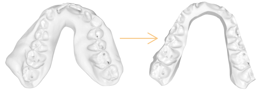
- Made in Centric Occlusion out of PVS or vax
- Vestibular part is trimmed to the middle of the vestibular cusps
- Lingual part is trimmed as much as possible but within stabilizing rigidity of the template
Correct Bite Template

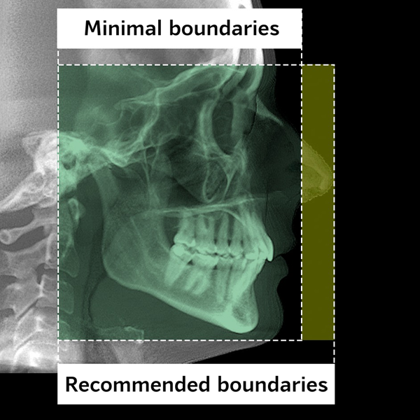
WHICH CBCT SCAN CAN BE USED TO MAKE A REPORT
-
- Voxel size no more than 0.3(0.25 or less is better)
- The patient must be in a natural bite!The use of a bite plate is prohibited. If the patient has a bite template, its use is mandatory!
- Minimum boundaries:Upper – no lower than the middle of the frontal sinus
Lower – the chin must be in the entirety
Front – preferably the tip of the nose should be in
(but can be trimmed if the TMJ does not fit)
Posterior – TMJ
All anatomical structures of the joint must be included: all walls of the fossa and the lateral pole of the head
-
- Voxel size no more than 0.3 (0.25 or less is better).
- The patient must be in a natural bite!
The use of a bite plate is prohibited. If the patient has a bite template, its use is mandatory! - Minimum boundaries:
Upper – no lower than the middle of the frontal sinus
Lower – the chin must be in the entirety
Front – preferably the tip of the nose should be in
(but can be trimmed if the TMJ does not fit)
Posterior – TMJ
All anatomical structures of the joint must be included: all walls of the fossa and the lateral pole of the head.
WHAT IS NEEDED FROM THE DIAGNOSTIC CENTER WHERE THE CBCT SCAN WILL BE DONE?
Mistakes in the making of the scan may result in the inability to perform some items in the report or in incorrect diagnostic data.
Therefore, we have prepared for you an instruction manual with scanning parameters, which you can download and give to the radiologist / lab technician.
You can always contact us by writing to WhatsApp or Telegram!
Therefore, we have prepared for you an instruction manual with scanning parameters, which you can download and give to the radiologist / lab technician.
You can always contact us by writing to WhatsApp or Telegram!
Important: diagnostic centres benefit from continuous cooperation with the doctor. Therefore, an instruction with requirements is normal.